Looking inside the brain with imaging technology
Brain scans have enabled scientists to learn more about Alzheimer’s than ever before1
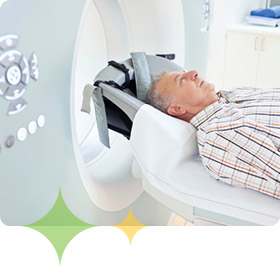
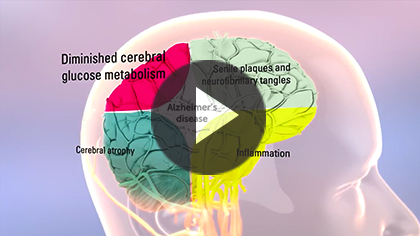
 Scientists can now see inside the brain with exquisite detail using a range of brain imaging techniques. Most often, these techniques are used by doctors to help diagnose Alzheimer’s disease. Brain imaging techniques are also used to rule out other conditions that may cause similar symptoms to Alzheimer’s disease, but require different treatments.
New imaging technologies are also being investigated to see if they may be able to help detect Alzheimer’s disease at a very early stage.
Imaging techniques used in Alzheimer’s disease research1
- Structural imaging – Provides information about the shape, position, or volume of brain tissue. These techniques include magnetic resonance imaging (MRI) and computed tomography (CT)
- Functional imaging – Reveals how well cells in different areas of the brain are working by showing how actively they use glucose (a type of sugar) or oxygen. These techniques include positron emission tomography (PET) and functional MRI (fMRI)
- Molecular imaging – Uses highly targeted radiotracer (a chemical compound that has a small, safe amount of radioactivity incorporated into it) to detect changes in cells associated with disease. For example, there are now radiotracers that can highlight beta-amyloid protein in the brain during a PET scan. These techniques include PET, fMRI, and single photon emission computed tomography (SPECT)


