Diminished cerebral glucose metabolism in Alzheimer’s disease
Diminished cerebral glucose metabolism (DCGM) is a key underlying pathology in the Alzheimer’s brain1
Cerebral atrophy, inflammation, amyloid plaques, and neurofibrillary tangles are characteristic pathological features in the Alzheimer’s brain, and they are not directly addressed by current drug treatments.2,3
Now, extensive evidence from three decades of research has established another pathological feature – DCGM, also known as glucose hypometabolism.1
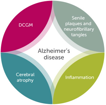
DCGM is a crucial aspect of Alzheimer’s disease that cannot be addressed with drug treatments1,3
Understanding of how cerebral glucose metabolism changes during aging and Alzheimer’s disease has grown rapidly over the past three decades. From the now-extensive scientific literature, it is clear that in Alzheimer’s disease:
- DCGM occurs in a characteristic pattern, commonly affecting the posterior cingulate, parietal, temporal, and prefrontal regions of the brain4-10
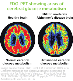
- Low glucose metabolic rates correlate with plaque density and lower cognitive scores11
FDG-PET showing areas of cerebral glucose metabolism
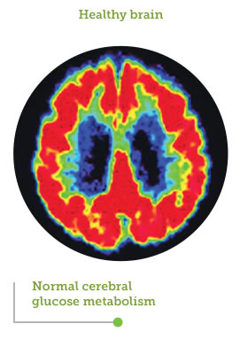
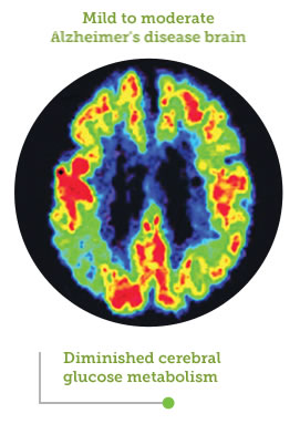
Image source: Small GW, Ercoli LM, Silverman DHS, et al. Cerebral metabolic and cognitive decline in persons at genetic risk for Alzheimer’s disease. Proc Natl Acad Sci USA. 2000;97(11):6037-6042. Copyright 2013 National Academy of Sciences, U.S.A.
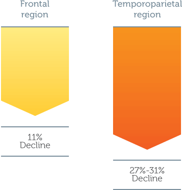
The brain’s capacity to utilize glucose as fuel is diminished by up to 25%, leaving a large portion of its energy needs unfulfilled1
DCGM occurs early9,10
DCGM occurs early in Alzheimer’s disease, particularly in people at genetic risk of developing the disease. The major genetic risk factor for late-onset Alzheimer’s disease is the carriage status of the epsilon 4 variant of the apolipoprotein E gene (APOE4 carriers).9,10
APOE4 carriers have been shown to have a low cerebral glucose metabolic rate, despite showing no signs of cognitive impairment or plaque deposition. This pattern of DCGM can be detected in young adult APOE4 carriers (mean age 30.7 years old), consistent with the notion that Alzheimer’s disease begins decades before cognitive impairment is evident.10
Brain areas showing DCGM in young adult APOE4 carriers10
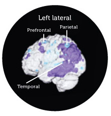
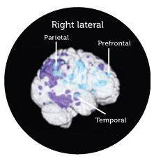
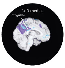
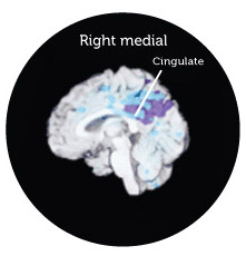
Adapted from Reiman et al. 2004. Three-dimensional surface projection map of abnormal DCGM in young adult APOE4 carriers superimposed on patients with probable Alzheimer’s disease mapped to a volume-rendered magnetic resonance image of the brain. There was a similar pattern in both groups, with notable decreases in the posterior cingulate, parietal, temporal, and prefrontal cortices.
In patients with probable Alzheimer’s disease, DCGM can be detected across genotypes12
DCGM is not exclusive to APOE4 carriers. In a study of patients with probable Alzheimer’s disease, DCGM was consistent across genotypes APOE3/E4, APOE3/E3, and APOE4/E4.12
DCGM in patients with probable Alzheimer’s disease, demonstrated with FDG-PET12
